

Mesenchymal stem cells have the ability to pre-differentiate into various tissues in order to regenerate them. In this particular case, they can be applied after being pre-differentiated into muscle progenitor cells, in combination with activated lymphocytes.
Initially, the cellular material controls the inflammatory process to stop damage and create an environment conducive to repairing the compromised tissue.
Repair is achieved by transforming the implanted stem cells into the target tissue: atrophied or injured muscles. A tissue repair process is then activated to repair the damage.
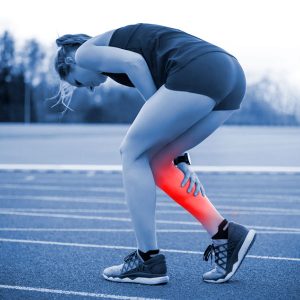
Various types of muscle damage or injury can be treated with cell therapy.
Some of these injuries cause fibrous scarring, which occurs when muscle tissue is replaced by fibrotic tissue. The greater the extent of fibrosis, the greater the functional consequences for the muscle. Fibrosis is associated with decreased muscle volume, signs of muscle atrophy in areas adjacent to the scar, loss of elasticity and contractile strength, and predisposes to further injury.
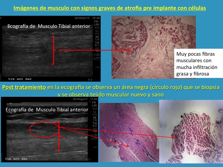
The clinical evidence presented shows the evolution of a tibialis anterior muscle with severe signs of atrophy before and after treatment. The initial histological study reveals a minimal presence of muscle fibers, displaced by a predominant infiltration of fatty tissue and fibrosis. Following the application of the cell protocol, ultrasound follow-up identified areas of regeneration that, when analyzed by biopsy, confirmed the formation of new, healthy, structured muscle tissue. This case documents the ability of cell therapy to reverse chronic degenerative processes and restore lost muscle architecture.
To prevent residual fibrosis and accelerate recovery times, cell therapy is an alternative that provides the material and specific inflammation needed for tissue to regenerate with the architecture and histology of healthy muscle, allowing contractile function and muscle elasticity to remain intact.
The healing time for a muscle tear can range from 3 to 16 weeks, depending on the extent of the injury. The healing of tears involves the regenerative capacity of muscle tissue and the formation of fibrous scarring.
Treatment seeks to stimulate regeneration to compete with scarring. The type of inflammation and the amount of raw material available to generate new muscle are essential for controlling fibrosis.
The use of muscle-differentiated stem cells with specific lymphocytes aims to provide the damaged tissue with the necessary raw material and inflammatory environment that prevents fibrosis and promotes the restoration of muscle structure.
There are different degrees and mechanisms of muscle injury.
The following cases are considered direct muscle injuries:
Indirect muscle injuries are the result of an intrinsic force generated by a sudden contraction of the muscle. These are commonly known as:
Muscle tissue accounts for between 40 and 45% of body weight. A muscle fiber is its basic structural element and is composed of a long structure of cells connected to the tendon or bone on which it acts. Muscles produce and modulate joint movement and are controlled by peripheral nerves.


Traditionally, muscle injuries—such as tears, severe contusions, and atrophy—have been treated using protocols focused on symptom control and physical rehabilitation. In the acute phase, standard treatment consists of cryotherapy, compression, and the use of analgesics and nonsteroidal anti-inflammatory drugs to limit edema and pain. While these methods stabilize the initial injury, their ability to influence biological tissue regeneration is limited, focusing primarily on inflammation management.
In cases of atrophy or more severe tears, conservative treatment is based on prolonged physical therapy, including progressive therapeutic exercises and neuromuscular electrostimulation to attempt to recover lost mass and functionality. However, in significant tears or chronic atrophy processes, scar tissue (fibrosis) and fat infiltration often form, compromising the muscle’s long-term elasticity and strength. In cases of complete rupture, surgery is the conventional option for tissue repair; however, this carries anesthetic risks and does not guarantee restoration of the original muscle architecture, sometimes resulting in incomplete recovery of pre-injury motor performance.